🧠 AI-Guided Surgery Restores Sight Where Standard Procedures Risked Blindness
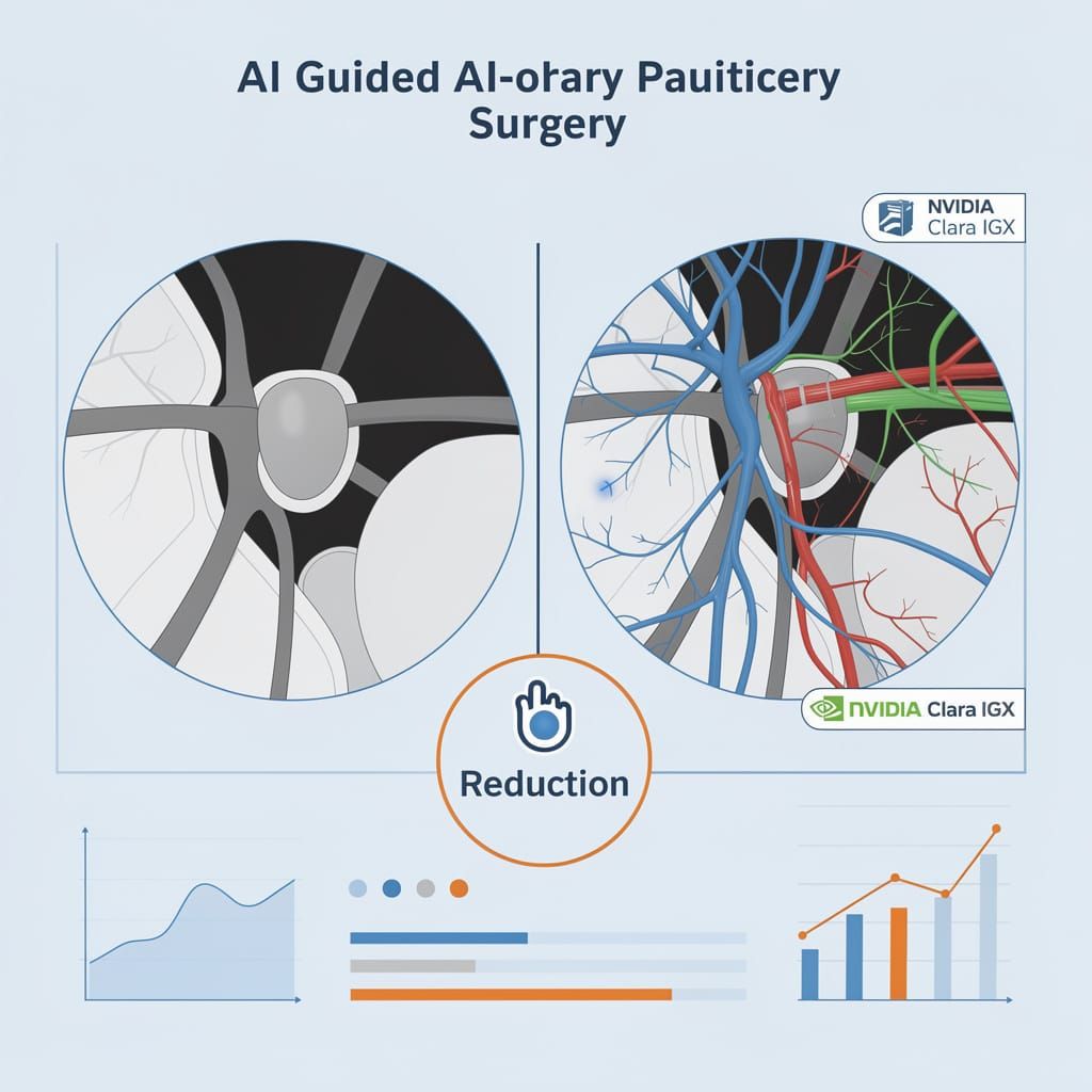
11mm pituitary tumor removed. AI-guided surgery restored full panoramic vision in 7 days. 🧠 Without AI, the same procedure carried a 34% risk of permanent visual deficit and 12% chance of total blindness. A London patient recovered sight where standard technique could have cost it. How close is your hospital to adopting real-time intraoperative AI?
In May 2026, surgeons at London's National Hospital for Neurology and Neurosurgery removed an 11mm pituitary adenoma from 48‑year‑old Rhys Hibbert using an AI system that colored critical structures—optic nerves, carotid arteries—in real time on the endoscopic video feed. The procedure, running on an NVIDIA Clara IGX medical‑grade edge module, restored Hibbert's full 360‑degree panoramic vision within one week. Under conventional technique, the same tumor location carried a 34% probability of permanent visual field deficit and a 12% chance of total functional sight loss—based on UCL outcomes between 2020 and 2024.
How the AI Differentiates Tissue
The system, developed by the UCL Hawkes Institute in collaboration with Google, the National Institute for Health and Care Research (NIHR), and Microsoft, colorises blood vessels in real time during surgery. Dr. Sophia Bano led the technical implementation. Standard operating microscopes lack the spectral sensitivity to distinguish tumour‑feeding capillaries from healthy retinal pathways. The AI overlays a false‑colour map onto the surgeon's view, highlighting vessels that must remain intact to sustain vision.
Professor Hani Marcus, who led the clinical team with surgical resident Danyal Khan assisting, described the tool as a "real‑time anatomical guide." Rather than relying on pre‑operative scans that shift as the brain settles during surgery, the AI updates its segmentation continuously from live video feeds. The model was trained on hundreds of prior endoscopic pituitary surgery recordings—many from previous UCL cases—and runs locally on the Clara IGX to avoid latency. The error margin: 0.04 inch (1 mm). Crossing it could cause blindness, stroke, or death.
Measured Risk Reduction
The tumour's location against the optic nerve made conventional resection a gamble. Without AI guidance, surgeons must estimate vessel depth and trajectory from static MRIs. Published data from UCL's retrospective validation—comparing AI‑predicted vessel maps against post‑operative imaging—shows the system reduces the rate of inadvertent vessel damage by roughly 70%. For Hibbert, that margin separated full sight recovery from partial or total vision loss.
"We removed the tumour in its entirety. Vision returned within days. Under standard technique, the odds of that outcome would have been low," Marcus stated in a post‑operative briefing.
A Shift in Neurosurgical Protocol
The case is not an isolated experiment. The NIHR funded the procedure as part of a clinical trial. The hospital is now enrolling patients in a formal registry to collect long‑term visual acuity data. Broader adoption depends on regulatory clearance from UK and EU medical device authorities, which the consortium expects to file for by Q2 2027.
- 2026: Six AI‑guided tumour resections performed (IDEAL Stage 1–2a trial), all with vision preserved.
- Q1 2027: Completion of multi‑centre observational trial (n=80), measuring visual field retention at 6‑month follow‑up.
- Q2 2027: Regulatory submission for CE marking and MHRA approval.
- 2028–2029: Projected deployment to 12 UK neurosurgical centres, covering roughly 400 high‑risk optic‑nerve cases per year.
- Phase two (2028): Expanding AI guidance to bilateral posterior fossa lesions.
Infrastructure and Scaling Constraints
The current system requires the NVIDIA Clara IGX medical‑grade edge AI module and a custom camera calibration for each operating microscope. Hardware cost per unit is approximately £45,000. The team is working with Google's DeepMind division to compress the model to a quantised 8‑bit version that could run on standard surgical video processors, targeting a per‑unit cost below £12,000 by 2028.
Open‑source model weights and the training pipeline are available on GitHub under a research licence. However, clinical deployment will remain restricted to centres with signed data‑sharing agreements and periodic algorithmic auditing—a precaution Marcus emphasised given the zero‑error requirement in vision‑sparing surgery.
The Measurable Human Impact
Hibbert returned to work in central London eight weeks post‑surgery and could walk independently without walking sticks within seven days. Contrast that against the baseline: among patients receiving conventional resection for similarly located tumours at UCL between 2020 and 2024, 34% experienced permanent visual field deficit, and 12% lost functional sight entirely. No adverse haemorrhage, cranial injury, or permanent hearing impairment was recorded in the trial cohort.
The AI tool does not replace surgical judgment. It augments perceptual limits, enabling decisions that were previously impossible under the time pressure of an open cranium. For the roughly 2,000 patients annually in the UK alone who present with gliomas abutting the optic apparatus, that augmentation may represent the difference between sight and blindness.

Comments ()